Lead Aprons and Thyroid Collars Are
NO LONGER NEEDED
During Dental X-Rays
OVERVIEW
WHAT ARE DENTAL X-RAYS?
Dental x-rays are used to make quick and painless images of your teeth and jaws. X-rays are invisible beams of energy, a form of radiation. The images are displayed on film or on the computer monitor (digital imaging) after the x-rays pass through an area of the body and are absorbed differently depending on the density of the structures. Dense body parts such as bones and teeth absorb much of the x-rays and will show up as white areas on the resulting image, while less dense body parts such as nerves and muscles absorb less, showing up as shades of gray.

INDICATIONS
WHEN SHOULD DENTAL X-RAYS BE OBTAINED?
Dental x-rays are used to diagnose diseases affecting the teeth and the bones since the inside of these structures is not seen when dentists look in your mouth. They provide important information to help plan the appropriate dental treatment.
They may be used to identify:
- Number, size, and position of the teeth
- Initial or advanced dental caries (a.k.a. tooth decay)
- Bone loss caused by periodontal disease (a.k.a. gum disease)
- Tooth infection
- Jaw fractures
- Problems of occlusion
- Jaw lesions
- Other teeth and bone abnormalities
FREQUENCY
HOW OFTEN SHOULD DENTAL X-RAYS BE TAKEN?
The type and frequency of dental x-rays depends on the patient’s needs which are determined based on the clinical exam and risk factors. If you are a new patient, dental x-rays may be requested to determine your oral health and to have a baseline to identify changes that may occur later.
How often dental x-rays should be taken depends on:
- Age and stage of development
- Present oral health and clinical findings
- Risk for dental caries and periodontal disease
- If you have any signs and/or symptoms of oral disease
TYPES OF DENTAL X-RAYS
There are different types of dental x-rays that can be taken with the receptor (film, sensor or plate) inside the mouth, called intraoral x-rays, or outside the mouth, called extraoral x-rays.
INTRAORAL X-RAYS
Periapical - evaluation of the crowns and the roots of teeth and the health of the bone surrounding the roots of the teeth
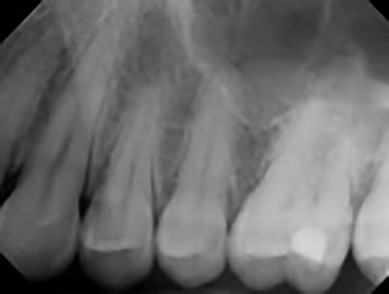
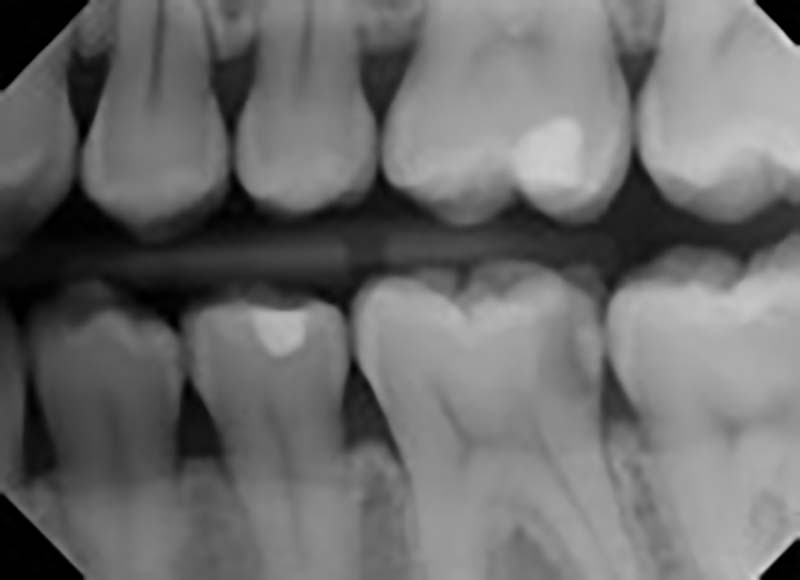
Bitewings – evaluation of the crowns of the upper and lower teeth for the presence of caries, the condition of existing fillings, calculus deposits, and the level and health of the bone surrounding the teeth
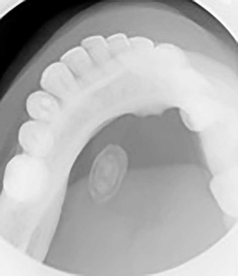
Occlusal – reveals the arch of teeth in the upper or lower jaw and is used to evaluate the effect of jaw lesions in the bone or to determine the presence of salivary stones in the floor of the mouth
EXTRAORAL X-RAYS
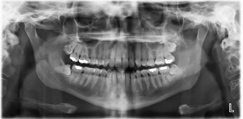
Panoramic- Shows all of the teeth including third molars (wisdom teeth), the maxilla, the mandible, the TMJs, and the maxillary sinuses
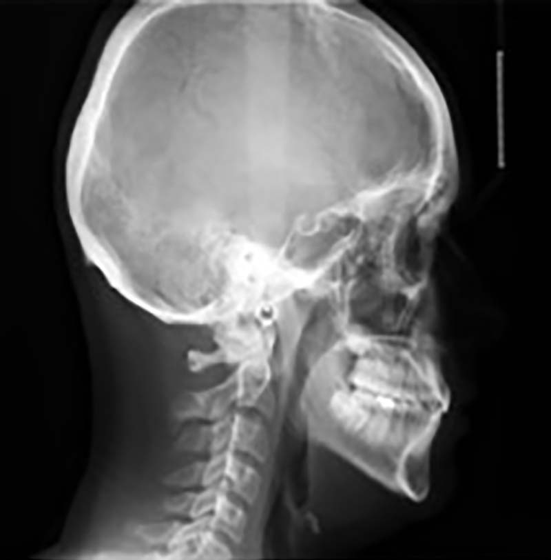
Cephalometric - Shows the entire head from a lateral or anterior view and is useful for orthodontic treatment planning to see the relationship between the teeth, the jaws, and the base of the skull
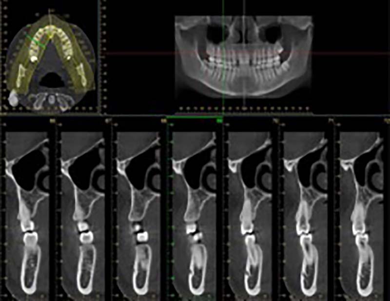
Cone Beam CT (CBCT) - Shows a 3D view of the teeth, the jaws, and the skull bones. It provides accurate dimensions of available bone for dental implants, accurate position of impacted teeth, and accurate location and extension of pathologies within the bones
RISKS - RADIATION EXPOSURE
WHAT ARE THE RISKS FROM RADIATION?
The risk from a single dental x-ray image is very small. However, some studies do show a slight increase in cancer risk, even at low levels of radiation exposure, particularly in children. To be safe, we do everything we can to keep the radiation exposure as low as possible.
ARE DENTAL X-RAYS SAFE? HOW MUCH RADIATION IS USED IN DENTAL X-RAYS?
We all are exposed to small amounts of radiation daily from the sun, soil, rocks, buildings, air and water. This type of natural radiation is called background radiation. The dentist will use only enough radiation necessary to evaluate the area of interest.
| Estimated effective dose (µSv) |
Equivalent amount of background radiation | ||
|---|---|---|---|
| Natural Background radiation | 8 | 1 day | |
| Airline passenger (4 hour flight) | 8 | 1 day | |
| Dental X-rays | 4 Bitewings | 10 | 1 day |
| Full mouth series (4 bitewings + 16 periapical | 35 | ~4 days | |
| Panoramic | 24 | 3 days | |
| Cephalometric | 6 | < 1 day | |
| Low-dose CBCT (Small FOV (3 teeth)) | 32-43 | 4-5 days | |
| Medium FOV (one jaw) | 113-136 | 14-17 days | |
| Large FOV (both jaws) | 269 | 34 days | |
| Medical | Chest X-ray (single view) | up to 10 | up to 1 day |
| Chest X-ray (2 views) | up to 100 | up to 10 days | |
| Head CT | up to 2000 | up to 8 months | |
| Chest CT | up to 3000 | up to 12 months | |
| Abdominal CT | up to 5000 | up to 20 months |
* Adapted from Ludlow et al. J Am Dent Assoc 2008;139(9):1237-43 and Ludlow et al. Dentomaxillofac Radiol 2015; 44: 20140197 based on conditions and equipment used at the UM School of Dentistry
The radiation used for dental X-rays has been compared to the amount of background radiation a person gets daily to help you understand how much radiation is given during the dental x-ray exam.
Comparison of Radiation Effective Dose from Various Dental and Medical Image procedures to Natural Background radiation (* adapted ref NCRP report No 177 and Image Gently)
DENTAL X-RAYS IN CHILDREN
There is very low risk from a single dental x-ray image. However, some studies shows a slight increase in cancer risk, particularly in children. Therefore, it is important to keep the radiation exposure as low as possible.
Dental X-rays For Children: What Parents Should Know
DENTAL X-RAYS AND PREGNANCY
Make sure to tell your dentist if you are pregnant. During your pregnancy, x-rays may be needed as part of your treatment plan for a dental disease. According to the American College of Obstetricians and Gynecologists, dental care, including dental x-rays, are safe during pregnancy. Use of the leaded apron and thyroid collar will protect you and your fetus from radiation exposure. Also, dental X-rays do not need to be delayed if you are trying to become pregnant or are breastfeeding.
PREVENTION
HOW CAN WE REDUCE THE RADIATION?
To make sure the child exposure is reduced as much as possible the Image Gently campaign recommends dentists to:
- Take x-rays based on the specific patient/ child’s needs and not as a routine test
- Use up to date equipment and techniques
- Use thyroid collars (to protect the thyroid gland that is more sensitive to radiation) and protective shields as needed for other body parts
- Use child-size exposure times
- Take Cone-Beam CT scans only when necessary
QUESTIONS
You should first talk to your dentist to discuss any concerns and questions regarding dental x-ray exams. For additional questions; you can visit the Image Gently website.
REFERENCES
What Parents Should Know about the Safety of Dental Radiology.

